The Shoulder

The shoulder is one of the most complex joints in the human body. What makes it remarkable is that it has minimal constraints, allowing us an incredible amount of motion. In fact, the shoulder is the most mobile joint in the human body. However, this motion also comes with a cost, as the very mechanisms that allow us so much motion also can be prone to injury.
If you're experiencing shoulder pain, call one of our offices or book an appointment online to get started.

Shoulder Anatomy
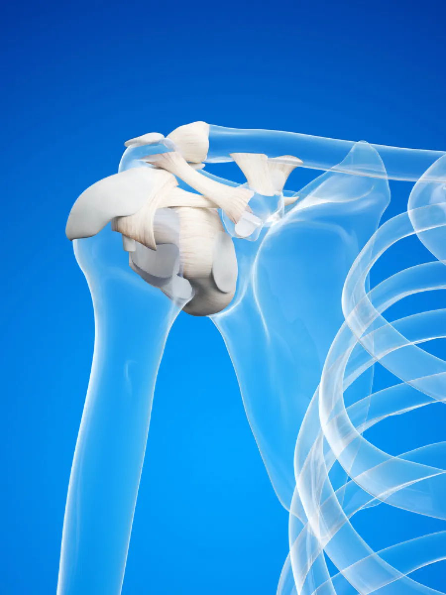
The shoulder consists of one major joint called the glenohumeral joint and a smaller joint called the acromioclavicular joint, or AC joint. The glenohumeral joint is a loose ball and socket joint which is what allows most of the motion of the shoulder. The glenoid is the socket portion of the joint, but it is very shallow. This socket is deepened by a cartilage structure known as the labrum. The labrum is what is responsible for much of the stability in the shoulder.
The AC joint connects the clavicle and a portion of the shoulder blade called the acromion. It helps with a person's ability to raise their arm up overhead. This joint is stabilized by 2 sets of ligaments, the acromioclavicular (AC) ligaments and the coracoclavicular (CC) ligaments.
A healthy functioning shoulder not only requires these joints to be in good condition, but also benefits from soft tissue support from muscles, ligaments, and tendons. The rotator cuff is one of the most important of these stabilizers in the shoulder. It is actually made up of 4 muscles all of which attach as tendons to the proximal humerus of the shoulder.

What causes shoulder pain?
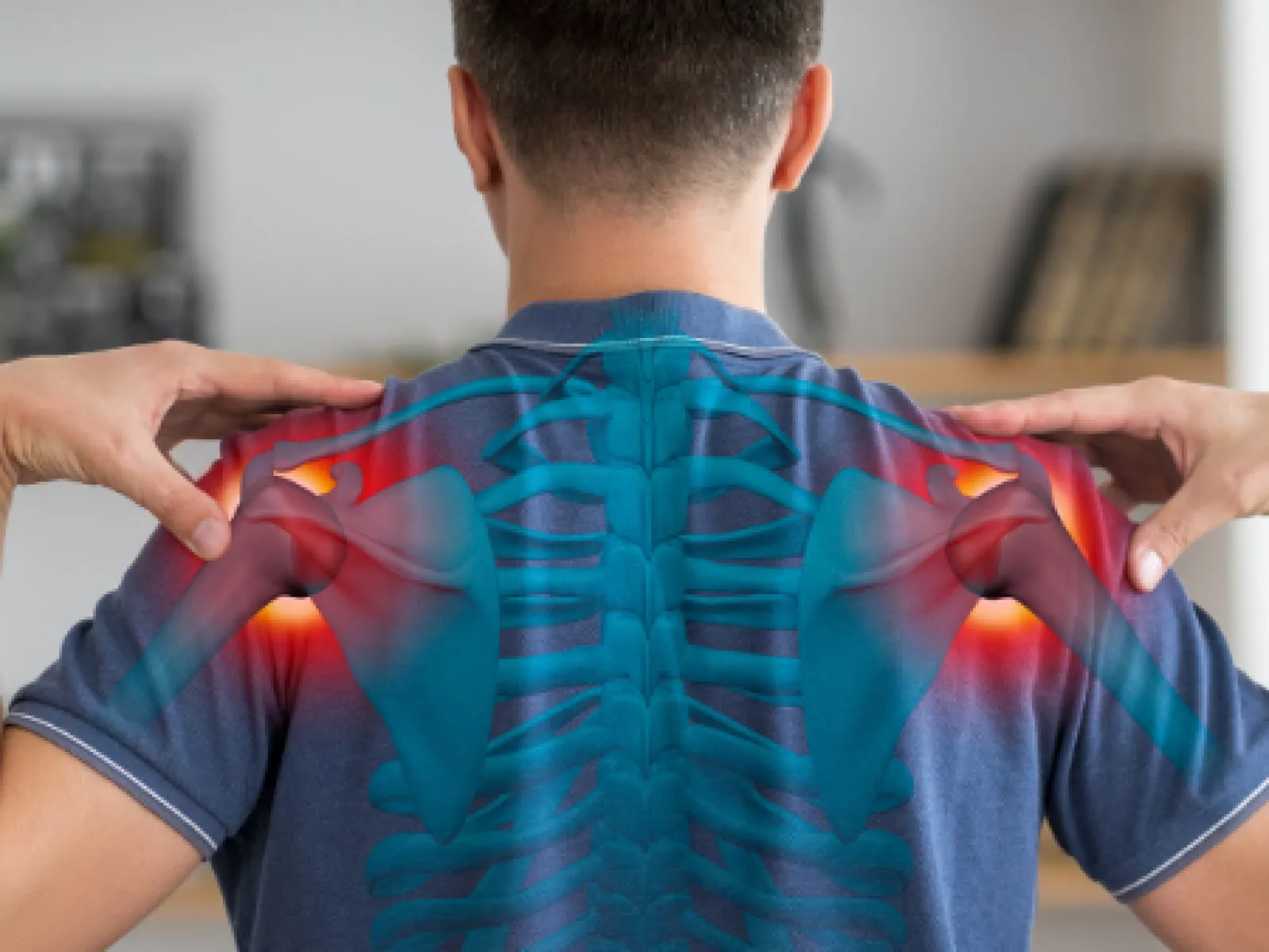
There are many causes of shoulder pain. Some common ones can be damage to the cartilage of the shoulder leading to arthritis, tears of the labrum caused by shoulder dislocations, tears of the rotator cuff, or inflammation known as shoulder bursitis. Pain from these injuries can be experienced in various locations, including along the shoulder blade, at the front of the shoulder, or at the top of the shoulder depending on the cause of your symptoms.
Common Conditions
Rotator Cuff Injury
Rotator cuff injuries are one of the most common causes of shoulder pain. Injuries can include partial or complete tearing of the tendons of the rotator cuff. Tears can affect a single rotator cuff tendon, or in more severe injuries multiple tendons can be affected. In younger patients traumatic injury, such as a fall from a height, can lead to rotator cuff tears. In older patients, rotator cuff tears are more often associated with chronic wear and tear that comes from daily use or from job related repetitive motions.
Symptoms of a rotator cuff tear include shoulder pain that worsens with activity as well as, in some cases, pain at night that can wake a person from sleep. Depending on the size of the tear, there can be accompanying weakness, especially with lifting the arm up overhead. In some cases this weakness can actually lead to loss of activity or range of motion of the shoulder.
Rotator cuff injuries are diagnosed by a combination of physical exam, X-rays, and when necessary, an MRI to look at the tendon itself. Depending on the severity of the tear, some can be treated with physical therapy and cortisone injections while others go on to require surgical repair.
Shoulder Impingement or Subacromial Bursitis
Shoulder impingement is one of the most common causes of shoulder pain. It is caused by inflammation in the fluid filled sac that sits on top of the rotator cuff. This inflammation is thought to be caused by compression of the rotator cuff between the humeral head and the underface of the acromion, a bone that is part of the scapula.
Symptoms usually occur without an injury event and often develop slowly over time. Pain occurs with overhead activity and reaching behind the back because in those positions the top of the shoulder blade rubs or impinges on the rotator cuff tendons. Loss of strength is not commonly associated with subacromial bursitis. Similarly, although people often experience pain with certain motions, the overall range of motion of the shoulder is usually maintained.
Shoulder impingement is diagnosed primarily through physical examination and X-ray. In some cases an MRI can also be ordered, but it is not usually necessary. The vast majority of patients improve with a combination of anti-inflammatories, physical therapy, and cortisone injections. Very rarely, surgery can be indicated in patients who do not have complete resolution of their symptoms.
Frozen Shoulder or Adhesive Capsulitis
Frozen shoulder is characterized by unexplained loss of range of motion in the shoulder. It is more common in women and most common between the ages of 40 to 60. It is not usually related to a traumatic injury, instead it tends to happen slowly over time. For reasons not entirely understood, a spontaneous inflammatory process in the shoulder leads to thickening of a structure called the capsule. This thickening and inflammation leads to reduced ability to move the shoulder and significant pain. There is an association between adhesive capsulitis and both diabetes and thyroid disorders.
Symptoms usually begin with generalized shoulder pain that then slowly leads to noticeable loss of motion in the shoulder. Initial stages are characterized by more pain, especially at night, while later stages are characterized by less pain and more significant limitations in overall motion. Motion can get so limited that it can severely impact a person's ability to engage in activities of daily living.
Diagnosis is primarily done with a physical exam. X-rays and MRIs are not usually as helpful in cases of adhesive capsulitis. Treatment is with cortisone injections into the shoulder joint, oral anti-inflammatory medications, and physical therapy. This is sufficient to treat the majority of cases, but when symptoms persist for more than 3 months, surgery is indicated to release some of the thickened capsule in the shoulder.
Shoulder Dislocation
Dislocations are very traumatic events. In the shoulder they are usually caused by a fall on an outstretched hand or by forced bending back of your shoulder such as occurs sometimes in contact sports. When the humerus bone comes out of its normal position and gets stuck, a dislocation occurs. This will usually cause severe pain and inability to move the shoulder. The shoulder can dislocate forward, backward, or downward, but the most common dislocation is when the upper arm bone, or humerus, moves forward and out of the socket.
In the event of a dislocation, especially if it is your first dislocation, the most important thing is to relocate your shoulder. Relocation will very quickly relieve the substantial pain a person experiences and improve their ability to move the shoulder. A shoulder dislocation is diagnosed on X-ray, and its subsequent relocation is confirmed on X-ray as well. Following a dislocation, a sling is used for a short period of time while the shoulder begins to heal. Physical therapy is then used to help regain range of motion and strength/
Shoulder Instability
Shoulder instability is one of the most common shoulder conditions. In general it is a sequelae of an initial traumatic shoulder dislocation. The trauma from dislocation can lead to tears in the labrum which is a cartilage bumper that surrounds the socket. Once someone has a torn labrum, there is a deficiency in the socket portion of the ball and socket joint. This can then lead to instability. In the worst cases, instability can be so bad that it can lead to future dislocations recurring more frequently.
AC Separations
AC Separations, more commonly known as shoulder separations, actually do not involve the main glenohumeral shoulder joint at all. Instead, they involve tearing of the ligaments around the end of the collarbone, or clavicle, which can compromise the acromioclavicular joint of the shoulder. AC separations are almost always caused by trauma, and usually from a direct force or blow onto the outside of the shoulder. Falls off of a bike or snowboarding falls are common causes. Depending on which ligaments tear, the clavicle usually is elevated when compared to the opposite shoulder. This leads to pain as well as noticeable deformity at the end of the collarbone.
Diagnosis of this injury is made with X-ray. A comparison to the uninjured shoulder will show exactly how elevated the collarbone is. The amount of displacement is measured and that is used to determine the grade of the AC separation. Grade I and II injuries are usually treated without surgery in a sling. Higher grade injuries may need surgical reconstruction. In these cases a small piece of cadaver tendon is used to reconstruct the ligaments around the collarbone, allowing the surgeon to recreate the original anatomy and bring the clavicle back into place.
Arthritis
Osteoarthritis is the most common type of shoulder arthritis. It can affect both joints in the shoulder, the large glenohumeral joint and the much smaller acromioclavicular (AC) joint. Osteoarthritis is usually caused by age along with wear and tear from activity and work. As such, it usually develops slowly over time and is most common in patients over 60 years of age. Progressive loss of cartilage leads to pain and joint stiffness.
There are other causes of arthritis. Some inflammatory conditions such as rheumatoid arthritis and psoriatic arthritis can also lead to loss of cartilage in the joint. Additionally, prior injury, such as dislocation or fracture, can change the biomechanics of the shoulder or permanently injure the cartilage leading to something called post-traumatic arthritis.
Symptoms usually begin very gradually with only a slight increase in pain or subtle loss of motion in the shoulder. Sometimes an unrelated injury in the shoulder can make underlying arthritis suddenly symptomatic as well. These symptoms tend to worsen over time especially as people get older. More severe arthritis can be associated with significant loss of motion as well as catching or cracking noises known as crepitus.
Shoulder arthritis is diagnosed primarily on X-ray, as this best shows the extent to which the joint has worn out. Once diagnosed, the treatment for arthritis depends on its severity. For more mild cases, preservation of range of motion and strengthening with physical therapy is very helpful. Significant flares of pain can sometimes also be controlled with cortisone injections. In severe cases, shoulder replacement can be a very effective option for restoring full range of motion and eliminating pain.
Fractures
Fractures in the shoulder joint can occur in multiple locations. They can be the result of a simple fall or from more high energy mechanisms such as car accidents or contact sports injuries. In the shoulder region the two most common locations that fractures occur are to the collarbone (clavicle fractures) and to the upper portion of the humerus bone (proximal humerus fractures).
All of these injuries should be first evaluated by a surgeon. The extent of the fracture can be evaluated on X-ray. Once the positioning and extent of the fracture are understood, a treatment plan can be put into place. Non-surgical treatment can involve immobilization in a sling for a period of time while the break heals. Surgical treatment will often involve placing a combination of plates and screws at the site of the fracture in order to stabilize the bone and allow for good future outcomes and function.
SLAP Tears
Superior labrum from anterior to posterior (Slap) tears are uncommon injuries to the shoulder. This socket is deepened by a cartilage structure known as the labrum. The labrum is what is responsible for much of the stability in the shoulder. These injuries can happen to anyone but most commonly occur in throwing athletes or in someone who fell on an outstretched arm. Symptoms often aren't noticeable right away and usually worsen with time. Typically, symptoms include a vague deep shoulder pain, weakness, or a popping or clicking sensation in the shoulder. A physical exam is used to diagnose this injury and oftentimes an MRI is ordered to confirm the diagnosis.
First line treatment for SLAP tears involves rest, physical therapy, anti-inflammatory medications, and, occasionally, a cortisone injection. In patients who are not improving or unable to fully return to their sport, an arthroscopic debridement and possible surgical repair of the labrum and bicep tendon may be considered.
Biceps Tendinitis or Ruptures
The Biceps tendon is a tendon that attaches to the anterior portion of the shoulder and allows for flexion and movement of the upper extremity. Biceps tendonitis is a repetitive stress injury caused by overuse of the arm and causes pain in the front part of the shoulder. A physical exam is used to diagnose biceps tendonitis. Treatment typically begins conservatively with physical therapy, anti-inflammatories and rest.
Biceps ruptures occur when the tendon gets torn off of its insertion point in the shoulder. This can occur in younger athletes who do heavy lifting with the shoulder or spontaneously in older patients. Symptoms include acute pain, decreased range of motion of the shoulder, swelling, bruising, and often a noticeable deformity in the arm. A diagnosis is made with a physical exam and typically an MRI is ordered to confirm it. Treatment depends on age and activity level and must be evaluated by a surgeon. Surgical fixation or non-surgical treatment with physical therapy may be recommended.
Calcific Tendonitis
Calcific tendonitis is inflammation and irritation of the rotator cuff tendons that, over time, can calcify and cause degeneration of the tendon. It is most commonly associated with an anatomical variant called subacromial impingement that causes chronic irritation of the rotator cuff tendon. This usually happens without any shoulder injury, instead people notice pain, decreased range of motion, or a clicking or catching sensation over time. X-rays will commonly show the calcifications, and in severe cases an MRI may be ordered to view degeneration or tears that may have occurred to the rotator cuff tendon.
Treatment involves physical therapy, rest, anti-inflammatory medications, and, commonly, a cortisone injection. Other newer treatments such as shock wave therapy may be considered in refractory cases. In patients with symptoms that are not improving or a tear in the rotator cuff surgical decompression of the calcium deposit may be considered.

Treatment
At Pacific Crest Orthopedics we customize a treatment plan to meet your individual needs. When it comes to the shoulder, some of the most common treatments can include different types of injections, use of anti-inflammatories, physical therapy, and use of a sling. For some conditions, surgery is indicated as the best option. See more on our surgery treatments.
Ephraim Dickinson, MD at Pacific Crest Orthopedics have extensive experience treating your shoulder pain and providing therapies to prevent future problems and to keep you active and healthy. Don't continue to suffer from shoulder pain or immobility. If you have questions about shoulder pain, call Pacific Crest Orthopedics in San Francisco or use the online booking tool to schedule an appointment today

